| Section categories |
|
Related Subjects [38]
This category includes brief overview of all related subjects.
|
|
Defining BioInformatics [7]
In this section we tried to briefly explain what bioinformatics is ?
|
|
Unviersities [30]
This contains information about universities that are offering bioinformatics degree programs.
|
|
Resources [24]
Contains information about bioinformatics resources including databases, tools and techniques.
|
|
Algorithms [31]
This category includes some of the basic algorithms that are usually used by bioinformaticians.
|
|
| Statistics |
Total online: 1 Guests: 1 Users: 0 |
|
Home » 2011 » June » 19 » Membrane Structure
9:37 PM Membrane Structure |
An Electron microscopic view of membranes
Surface specializations
Membrane specializations: Junctions
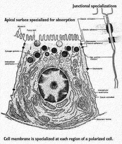
The drawing is of a polarized cell. The top is specialized for absorption and the bottom for transfer of materials to the blood stream. The sides have specialized junctions that keep the nutrients from entering the space between the cells. For more information about the thick filaments associated with these junctions, please consult the intermediate filaments web page. This figure was modified from Bloom and Fawcett, A Textbook of Histology, Chapman and Hall, N.Y., Twelfth Edition, 1994, Figure 2-10
Tight Junctions One of these is called a tight junction or "occluding junction" (zonula occludens). This is shown as the top junction in the above drawing. At this site, membrane glycoproteins and associated "glue" bind the cells together like double-sided "strapping tape" The freeze-fracture/freeze etch view of this junction (shown below) illustrates the ridges in the plane of the exposed leaflet. These are the proteins that bind to the proteins from the adjacent cell. This figure was modified from Bloom and Fawcett, A Textbook of Histology, Chapman and Hall, N.Y., Twelfth Edition, 1994, Figure 2-11.
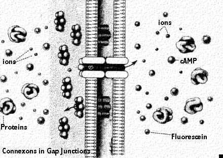
Gap Junctions Another type of junction allows communication between cells. This type is called a gap junction. Small molecules or ions can pass through, as we will see by the following figures.
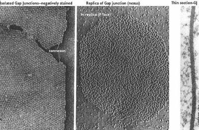
The above freeze-fracture /freeze etch image shows the internal view of the gap junction on the left. The proteins look like little donuts which reflects the fact that they are actually a channel. These proteins are "connexon" molecules. The side facing the cytoplasm (called the P face) is shown in the center panel. The region looks like aggregated lumps. Finally, the typical electron microscopic view is seen in the third panel. This shows a thin line between the two plasma membranes indicating a "gap junction". This figure was modified from Bloom and Fawcett, A Textbook of Histology, Chapman and Hall, N.Y., Twelfth Edition, 1994, Figure 2-14.
There are several ways to prove the cells are communicating by gap junctions. First, one can identify the connexon molecules by immunocytochemical labeling. Second, one can identify the actual junctional complex with freeze-fracture/freeze etch. To see if they are functional, however, one needs to inject one cell with a dye and watch to see if it is transferred to another cell.This cartoon diagrams a view of a gap junction showing molecules that can freely pass. Ions pass and in this way the cells can be electrically coupled together. Other small molecules that pass through include cyclic AMP (a second messenger) and the dye marker fluorescein. This last compound enables the scientist to study transport through the gap junction. This figure was modified from Bloom and Fawcett, A Textbook of Histology, Chapman and Hall, N.Y., Twelfth Edition, 1994, Figure 2-15.
|
|
|
Category: Related Subjects |
Views: 1445 |
Added by: Ansari
| Rating: 0.0/0 |
|
|
|