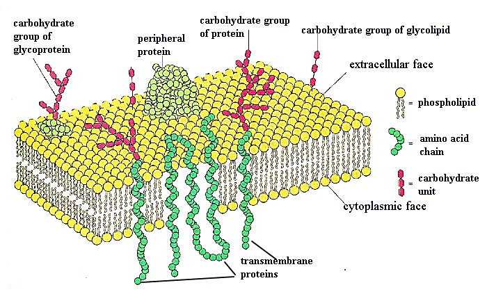
As you study different organelles, you will learn about important membrane proteins that function for that particular organelle. Transmembrane proteins are amphipathic, in that they have hydrophobic and hydrophilic regions
that are oriented in the same regions in the lipid bilayer. Another name for them is "integral proteins". Other types of proteins may be linked only at the cytoplasmic surface (by attachment to a fatty acid chain), or at the external cell surface, attached by a oligosaccharide. Or, these non-transmembrane proteins may be bound to other membrane proteins. Collectively these are called "peripheral membrane proteins" .
We will be studying specific membrane proteins in later lectures (ion channels, proteins in endoplasmic reticulum, etc). Therefore, this presentation will not spend much time on them. Review pp 486 and 487 in your text for information on insertion of a transmembrane proteins. Proteins inserted once through the membrane are called "single-pass transmembrane proteins." Those that pass through several times are called "multipass transmembrane proteins". They form loops outside the membrane Later lecturers will spend more time on this as key proteins are introduced.
The following figure shows transmembrane proteins passing through the lipid bilayer. Can you find the multipass and single-pass transmembrane proteins in this figure? The figure below is from Wolfe S.L., Molecular and Cellular Biology, Wadsworth Publishing Company, 1993.
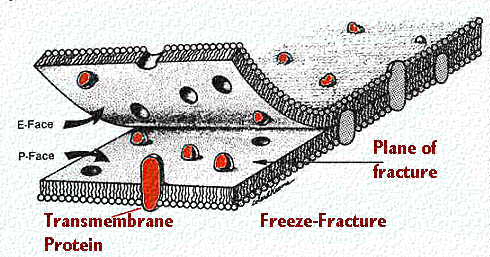
An EM view of membranes via freeze fracture/freeze etch .
You can best see protein distribution via a technique called freeze fracture/freeze etch. The freeze-fracture/freeze etch technique starts with rapid freezing of a cell. Then the frozen cells are cleaved along a fracture plane. This fracture plane is inbetween the leaflets of the lipid bilayer , as shown by this cartoon. The two fractured sections are then coated with heavy metal (etched) and a replica is made of their surfaces. This replica is then viewed in an electron microscope. One sees homogeneous regions where there was only the exposed lipid leaflet (Is the exposed surface made of polar or nonpolar groups? This figure was modified from Bloom and Fawcett, A Textbook of Histology, Chapman and Hall, N.Y., Twelfth Edition, 1994, Figure 1-3. Consult the section on Membrane Architecture for the answer.)
In certain areas of the cell, one also sees protrusions or bumps. These are colored red in the cartoon. Sometimes one can see structure within the bumps themselves. These are the transmembrane proteins.
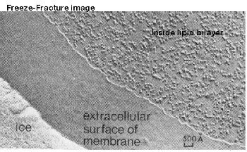
The following illustration will show you a freeze-fracture/freeze etch view. The organization or structure of the transmembrane proteins can often be visualized.