Bannykh, S and Balch WE Membrane Dynamics at the Endoplasmic Reticulum-Golgi Interface. J Cell Biol 138: 1-4 (1997)
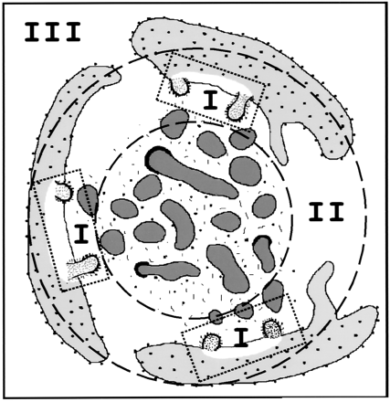
Tier I shows budding from ER that is arranged facing a central zone at one end of the Golgi complex. These buds become vesicles and are coated with COPII protein coats.
Tier II The ER faces a central zone called a vesicular-tubular cluster (VTC). After they lose their COPII coat, they merge with the VTC's carrying the soluble and membrane proteins to the Golgi complex.
Tier III illustrates the entire complex which is unique in the cytoplasm. It is termed the 'export complex' and contains unique proteins that suggest it is specialized for information flow to and from ER and the Golgi complex.
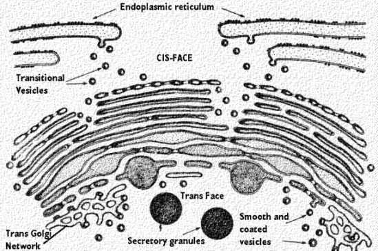
The above drawing shows an actual interface between the ER and the Golgi complex. The "Export complex" is seen at the top of the drawing. Note that the vesicle are moving to contribute to the cis-Golgi network of vesicles and cisternae.
The movement of these special transport vesicles is an energy requiring process. If one blocks production of ATP, the transport will not happen. This drawing shows how the rough endoplasmic reticulum forms vesicles (without ribosomes attached) that carry the newly synthesized proteins to the Golgi complex.
The inside of the vesicle becomes continuous with the inside of the Golgi cisternae, so that protein groups pointing towards the inside, could eventually be directed to face the outside of the cell.
Carbohydrate groups are attached and any subunits may be joined in these cisternae. The protein is then passed to the final region of the Golgi called the "trans face". There it is placed in vacuoles that bud from this region of the Golgi complex. These may be a certain size or density, characteristic of the cell itself. The vacuoles continue to condense the proteins and the final mature secretory granule is then moved to the membrane for secretion.
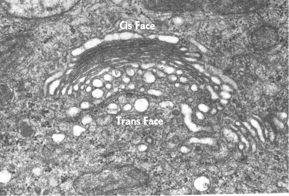
This electron micrograph illustrates a Golgi Complex. It is curved with its Trans face pointing away from the nucleus toward the cell periphery. The numerous vesicles in the area are transporting the proteins to and from cisternae.
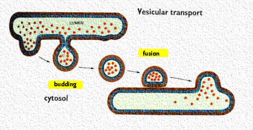
Transport of material in and out of the Golgi complex involves budding and fusion of vesicles. This cartoon shows that the membranes of each join and align themselves during the process so that the inside face remains in the lumen and the outside face remains towards the cytoplasm.